Substituting the canine with a premolar, moving the premolar with braces
If you've been through the phase of making space, and then had the expose & bond & traction not work out,
you are left with a visible gap. Extracting the canine will leave a gap where it was, and missing bone in the
area above the gap. The arc is incomplete and can't function as intended.
You can go the graft and implant route, or in some cases the choice is to move all the teeth up one step, so the premolar takes the place of the canine.
'Substitution of impacted canines by maxillary first premolars: A valid alternative to traditional orthodontic treatment' (1). In the case described no effort was made to erupt the canines because of their position, and because of 'dentoalveolar bioprotrusion' (no idea what that is) and other problems. The canines were extracted, as well as the mandibular second premolars. The premolars were moved forward and modified a bit, and the photos of the smile looks pretty OK after 3 years and 7 months.
Dr Becker writes:
But what if the long-term prognosis of the canine is poor because of its initial intractable position or as a
consequence of its having been through the processes of and orthodontic alignment over the period of many
months or years that were spent in its meticulous alignment? In such cases, perhaps it would have been
better to remove that particular canine at the outset and to have brought the first premolar to its place. (p
138, The Orthodontic Treatment of Impacted Teeth)
I asked about autotransplantation in Canada, and got this response back from Dr Sylvain Chamberland.
I went back to the website of your son's treatment and found the ceph taken on july 15, 2014. I wounder why nobody sugestted an alternate treatment plan that would have involve extraction of the ankylose canine and 3 premolars. It is quite clear from the tracing that maxillary incisors are proclined by 5° (mx1-SN = 109,4°; norms 104°), lower incisors are proclined by 14° (IMPA = 104°; norm = 90°) and interincisal angle is decreased by 15° (1/1 = 115°; norm 130°). Extraction of the ankylosed canine and 3 premolars would have help to reduce the dentoalveolar protrusion and achieve normal class I occlusion without the burden of waitng the right age to get an implant in a cosmetic zone where it will always be a challenge to have smile esthetics. I doubt that an autogenous transplantation would be a viable option. I would like show you this exemple where the patient was told that the canine might be ankylosed and that extraction of 3 premolars was the best option. My concern is that your son is into orthodontic treatment for so long and closing the space os canine and premolar extraction means 12 to 18 more months of treatment. It is sad that nobody has thought about an alternate treatment plan earlier.
Plus d'informations ici :
https://www.sylvainchamberland.com/en/dentition-en/impacted-canines/mechanotherapy/#comment-67137
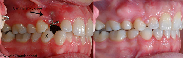
Photo from Dr Chamberland
The problem with a premolar as canine is described as:
If there is good contact between the lateral incisor and the first premolar then it has to be carefully
considered whether this should be accepted. The purists of occlusion will argue that the premolar is not
capable of providing good canine guidance. Aesthetically there are problems since the palatal cusp hangs
down. This can be disguised by grinding or rotating the tooth orthodontically so that the palatal cusp is
positioned more distally. The placement of a veneer on the premolar is another way of improving the
appearance.(2)